Figure 1.
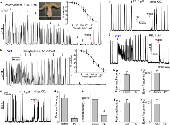
Phenylephrine inhibits the spontaneous and oxytocin-induced contractions in mouse uterus. (a) A sample trace shows the effect of phenylephrine (PE) on spontaneous contractility in a mouse uterine ring. The left inset shows a photograph of isolated mouse uterine horns and vagina. Yellow rectangles show the two segments of uterine horns typically used for isometric tension experiments. The dose-response curve for PE-induced relaxation is shown in the right inset (N = 8, the data were presented as mean ± S.E.). (b) A sample trace depicts that PE inhibits oxytocin-induced contractions in the mouse uterine ring in a concentration-dependent manner. The corresponding dose-response curve is shown in the inset (N = 6). (c) A sample trace shows the effect of 1 μM phenylephrine (PE) on a uterine ring isolated from CO2-anesthesized female mouse (N = 4). (d,e) Summary data for the frequency and amplitude of spontaneous contractions in uterine rings isolated from CO2-anesthesized female mice (N = 4). (f–k) Shown are sample traces and summary data for the effect of 1 μM phenylephrine (PE) on the frequency and amplitude of spontaneous and oxytocin (100 nM)-induced contractility in uterine strips from CO2-anesthesized female mice (N = 4). Active tension changes are shown in (a–c,f,g). The data were presented as means ± S.E. The value of “N” is the number of mice. The vertical arrows show the times where the drugs were added to the bath. OXT = oxytocin (100 nM).

