Figure 7.
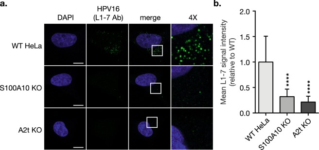
HPV16 capsid disassembly is dramatically inhibited in the absence of S100A10 and A2t. (a) WT, S100A10 KO, and A2t KO cells grown on chamber slides were treated with HPV16 pseudovirions (PsVs) (0.5 μg/1E6 cells) for 1 h at 4 °C to promote bulk binding. Cells were then washed and incubated for 7 h at 37 °C to promote endocytosis and intracellular trafficking. Cells were fixed with 4% PFA and immunostained with the anti-HPV 33L1-7 antibody (L1-7 Ab) that recognizes an internal epitope on the HPV capsid (green), marking disassembled capsid. Nuclei were stained with DAPI (blue) and a single confocal slice is shown in each representative image. Scale bar = 10 μm. (b) Quantification of L1-7 Ab signal intensity was measured as mean tonal intensity using Fiji. Results are shown as the mean ± s.d. (N = 15 images). Statistics: 1-way ANOVA with Dunnett’s multiple comparisons test – ****P < 0.0001.

