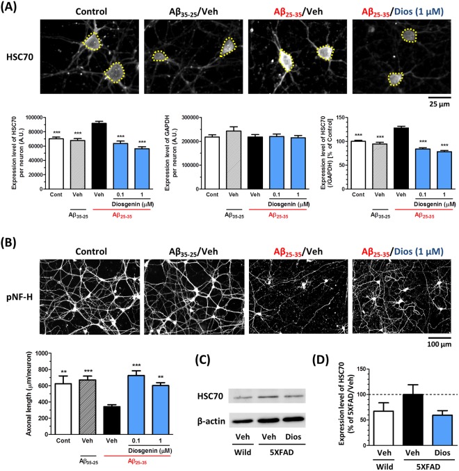
Figure 2.
Diosgenin treatment decreased expression of HSC70 in cultured neurons and in 5XFAD mouse brains. (A,B) Mouse cortical neurons (ddY, E14) were cultured for three days and then treated with Aβ35–25 (10 µM) or Aβ25–35 (10 µM) for three days. Then, neurons were treated with diosgenin (0.1 or 1 µM) or vehicle solution (0.1% EtOH) for four days. Neurons were fixed and immunostained for HSC70 and GAPDH, or pNF-H. (A) The expression level of HSC70 (yellow dotted line) and GAPDH in neurons were quantified, and the expression level of HSC70 (ratio to GAPDH) were quantified in each neuron. (B) pNF-H-positive axonal lengths were quantified for each treatment group. **p < 0.01, ***p < 0.001, two-tailed one-way ANOVA post hoc Dunnett’s test. (A) n = 115–217 neurons, (B) n = 11–15 photos were quantified for these analyses. (C,D) Wild-type and 5XFAD mice (Female, 7–8 months old) were treated with diosgenin (0.1 µmol/kg/day, p.o.) or vehicle solution (sesame oil) for 18 days. (C) WB for HSC70 in the wild-type mouse and diosgenin- or vehicle-administered 5XFAD mouse cortex. Representative cropped images are shown for each group. The full-length blots are presented in Supplementary Fig. 2D. (D) Quantitative value for the expression levels of HSC70 (ratio to β-actin), n = 3–4 mice.

