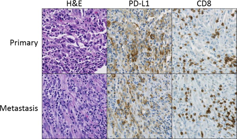
Figure 2. PD-L1 expression is concordant in paired primary and metastatic inflammatory myofibroblastic tumors.

Primary and metastatic specimens from three patients were included in the study and all were concordant for PD-L1 expression. Representative examples, including a primary inflammatory myofibroblastic tumor of the lung (row 1) and chest wall metastasis from the same patient (row 2), stained with H&E, PD-L1, and CD8 are shown. In both tumors, membranous PD-L1 expression is observed on tumor cells and is associated with CD8+ T cell infiltration. Original magnification 200×.
