
Figure 3. Mixed adaptive and constitutive PD-L1 expression in inflammatory myofibroblastic tumors.
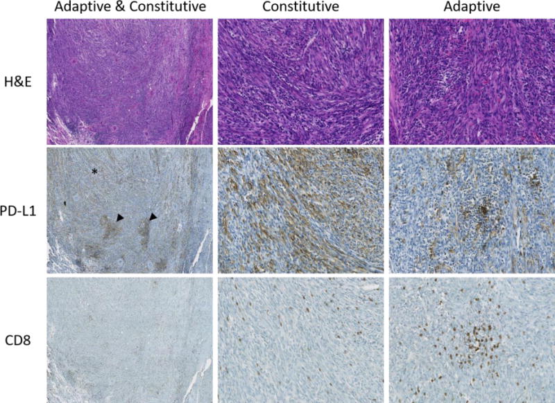
H&E, PD-L1, and CD8 stains of an inflammatory myofibroblastic tumor showing a combination of adaptive and constitutive expression. Heterogeneous PD-L1 expression can be seen at low power (first column), including diffuse constitutive expression (asterisk) and focal higher intensity adaptive expression (arrowheads). Higher magnification images highlight areas of constitutive expression (second column) and adaptive expression (third column). Original magnification 40× (first column) and 200× (second and third columns).