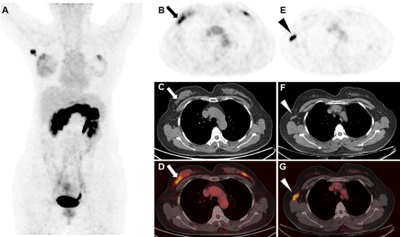
FIGURE 3.

An ER-positive breast invasive ductal carcinoma (arrow) with metastatic lymph node (arrow head) was clearly visualized in the right breast of a 47-year-old patient during the secretory phase of her menstrual cycle (A, maximum intensity projection; B and E, 68Ga-RM26 PET; C and F, CT; D and G, fusion images). The SUVmax of the right breast tumor, metastatic lymph nodes, and normal breast tissue were 4.47, 16.14, and 2.20, respectively. An occult lesion was also found on the left breast, but the patient rejected further surgery and the lesion is still under follow up.
