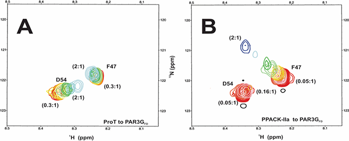
Figure 5:

2D 1H-15N HSQC NMR titrations of PAR3GFD (44–56) in the presence of ProT and PPACK-IIa. All NMR samples were in 25mM H3PO4, 150 inM NaCl, 0.2 mM EDTA and 10 % D2O (pH 6.5). (A) For the PAR3GFD binding studies with ProT, starting complexes included 37.5 μM PAR3GFD (44–56, 15N-F47, 15N-D54) in 70 μM ProT. The serial dilutions resulted in ProT to PAR3GFD ratios that spanned from 2:1 to 0.3:1. (B) For PPACK -IIa, starting complexes included 37.5 μM PAR3GFD (44–56, 15N-F47, 15N-D54) in 70 μM PPACK-IIa. The serial dilutions resulted in PPACK-IIa to PAR3GFD ratios that spanned from 2:1 to 0.05:1. Representative data sets are shown. Colors for the HSQC crosspeaks span from blue (highest protein-peptide ratio) to red (free peptide).
