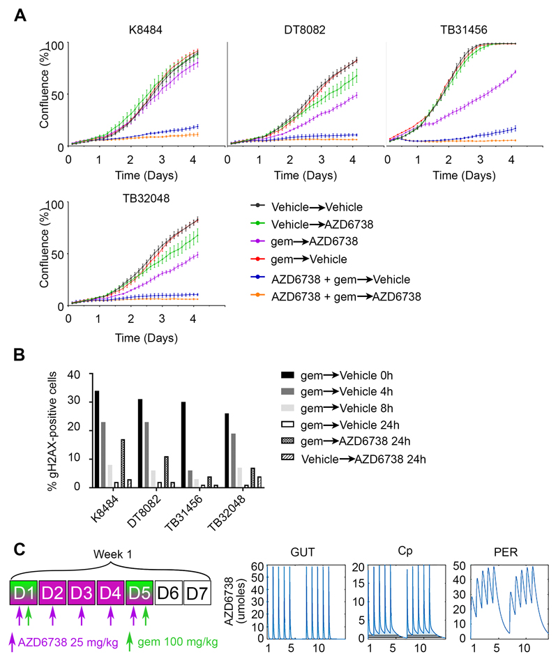
Figure 4. Scheduling of the combination of AZD6738 with gemcitabine.
A, Four cell lines from the KPC mouse model were plated at low density and treated with the first condition for 16 hours, then the medium was replaced with the second condition and cell growth was monitored by time lapse microscopy every 3 hours for 4 days. Drugs were used at the GI50 (72h) concentrations. B, Cells were incubated with 10 nM gemcitabine for 16h, then fixed at the time point shown after washout into fresh medium or with 300 nM ATRi. Immunofluorescence followed by quantitative microscopy was realized to obtain the percentage of cells positive for γH2AX. C, Dose-schedule schematic (left) of the in silico modelling of AZD6738 mouse PK for the gut, circulating and peripheral compartments (right).

