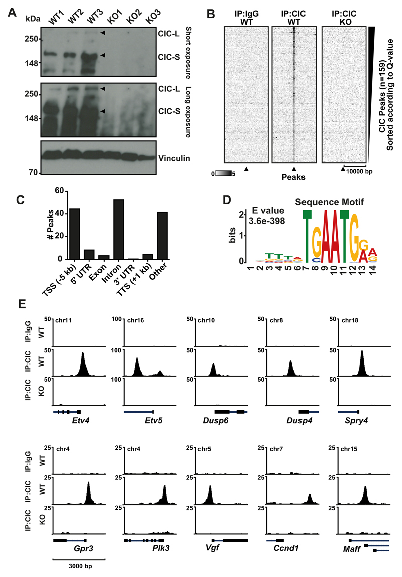
Figure 1. Genome-wide characterization of CIC targets in mESCs.
A) CIC Immunoblot of three WT mESC clones and three Cic-KO clones B) Heatmap at CIC peaks showing IgG and CIC in WT and Cic KO C) Motif analysis using MEME-ChIP. Top motif identified is depicted with corresponding E-value. D) Distribution of peaks according to gene elements. E) ChIP-seq tracks of CIC occupancy at selected target genes in WT and KO mESCs.

