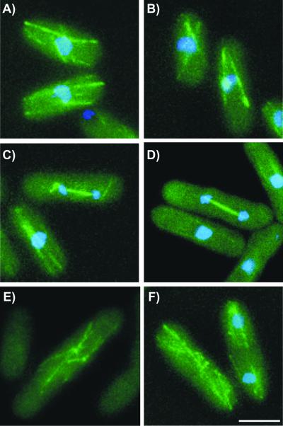
Figure 7.
Klp5p and Klp6p localize to cytoplasmic microtubules in interphase and spindles in mitosis. Klp5-GFP and Klp6-GFP were visualized in live cells (green) with DNA staining (Hoescht 33342, blue) by deconvolution microscopy. (A) Klp5p-GFP localized to cytoplasmic microtubules in interphase. (B) Klp6p-GFP localized to cytoplasmic microtubules in interphase. (C) Klp5p-GFP localized to a mitotic spindle in the nucleus, and to the “astral” microtubules in the cytoplasm (arrows). (D) Klp6p-GFP localized to a mitotic spindle in the nucleus. (E) Klp5p-GFP redistributes to the interphase microtubules as they are formed at the conclusion of mitosis. (F) Klp6p-GFP redistributes to interphase microtubules after mitosis. These first appear as the “post anaphase array” near the region of septation. Bar, 5 μm.

