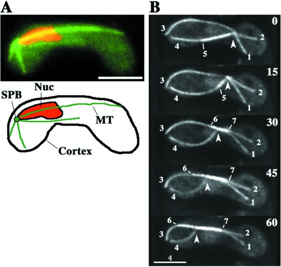
Figure 1.
Microtubule organization during nuclear oscillation in wild-type cells. (A) Wild-type cell (strain CRL152) expressing GFP-tagged alpha-tubulin in the nuclear oscillation stage. The cell was observed in a single focal plane. GFP-labeled microtubules and chromosomal DNA stained with Hoechst 33342 are shown in green and red, respectively. Bar, is 4 μm. (B) Projections from optical sectioning microscopy of a single cell in the nuclear oscillation stage. Each projection was constructed from images of 15 different focal planes (see MATERIALS AND METHODS). Barbed arrowheads indicate the position of a microtubule focus where the SPB is located. Large numbers indicate time in seconds. Small numbers identify individual microtubules. Bar, 4 μm.

