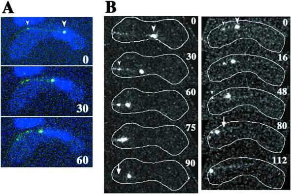
Figure 9.
Dynamics of GFP-tagged DHC in living meiotic cells. (A) Time-lapse series of a meiotic cell expressing GFP-DHC (green) and stained for DNA (blue) at a single focal plane. (B) Projections of meiotic cells expressing GFP-DHC. Each projection was constructed from images of a single cell at 15 (left column) or 8 (right column) different focal planes. Each column shows projections of a single cell. White lines indicate the cell cortex. Large arrowheads indicate large GFP dots, which are located at the leading end of the moving nucleus. Small arrowheads indicate small GFP dots located at the site where a GFP line contacts with the cell cortex. In B, cortical positions of some of the small GFP dots are obscured by the projection. Arrows indicate the sites where the small GFP dots disappeared. Numbers indicate times in seconds.

