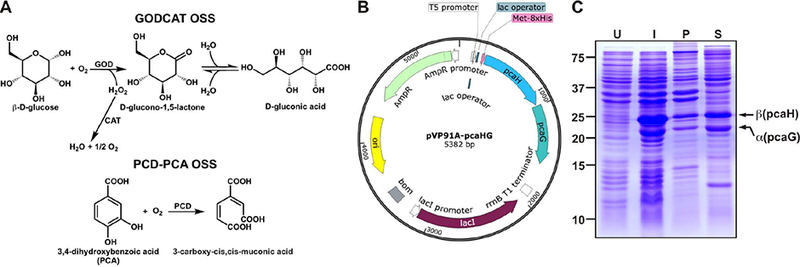
Figure 1. Recombinant expression of P. putida PCD in E. coli.

(A) Plasmid map of pVP91A-pcaHG containing the α (pcaG) and β (pcaH) subunits of PCD. The complete sequence of this plasmid is shown in Supplementary Figure 1. Locations of the T5 promoter, lac operator, and 8xHis are also shown. (B) SDS-PAGE analysis showing the whole cell lysate of uninduced (U), and induced (I) BL21 cells. Induction under optimal conditions resulted in a minor insoluble pellet (P) and a major soluble (S) fraction of PCD. The MW of α and β subunits are 22.4 kDa and 28.3 kDa respectively.
