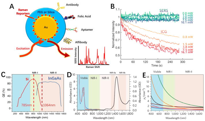
Figure 1.
(A) Schematic diagram of biocompatible surface-enhanced Raman scattering (SERS) nanoparticles that are encoded with Raman reporter molecules and conjugated with targeting ligands for in-vivo and intraoperative cancer detection. (B) Graphs comparing the photostability between SERS tags and indocyanine green (ICG) at various laser excitation powers. ICG is a common organic fluorophore used in fluorescence image-guided surgery. (C–E) Emergence of two windows in the near infrared (NIR) for in-vivo imaging and spectroscopic detection. (C) Sensitivity curves for typical cameras based on silicon (Si) or indium gallium arsenide (InGaAs), which are sensitive in the first and second near-infrared windows, respectively. The excitation wavelengths at 785 nm and 1064 nm are indicated by vertical dotted lines. (D) Absorbance of oxygenated (red curve) and deoxygenated (blue curve) hemoglobin in the visible and NIR spectrum, together with water absorbance (black curve) at 1400–1500 nm. (E) Plots of the scattering attenuation coefficient as a function of wavelength for various ex-vivo tissues (from top to bottom: green curve = brain tissue, yellow curve = intralipid tissue phantom, black curve = skin, brown curve = cranial bone, purple curve =mucous tissue, red curve =subcutaneous tissue, blue curve = muscle tissue). (B) and (D & E) were adapted with permission from references [4] and [10], respectively.

