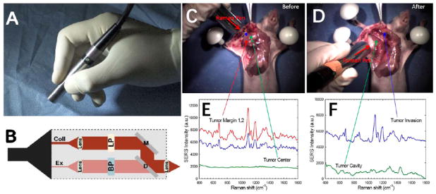
Figure 2.
(A) Photograph of a handheld spectroscopic device called the “Raman Pen” for high-sensitivity detection of tumor margins, metastatic lymph nodes, and micro-metastases during surgery. (B) Optical layout of the pen device: Ex = excitation fiber, Coll = collection fiber, BP = bandpass filter, LP = long-pass filter, D= dichroic mirror, M = total reflection mirror. (C–F) Demonstration of spectroscopic guided surgical resection of a tumor using SERS nanoparticle tags and a hand-held spectroscopic device. (C) Photograph of the anatomical locations where SERS spectra were obtained, shown in (E), across a xenograft tumor prior to resection. (D) Photograph of the anatomical locations where the SERS spectra were obtained, shown in (F), across the tumor cavity and surrounding areas after tumor resection. Note that scanning areas outside of the primary tumor location lead to a surprise finding of a satellite micrometastatic legion that would have otherwise gone unnoticed by the surgeon. (A&B) and (C–F) were adapted with permission from references [19] and [1], respectively.

