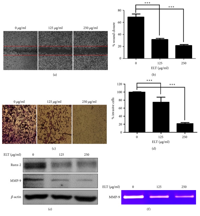
Figure 3.
Effects of ethanol extracts of Lycopus lucidus Turcz. ex Benth (ELT) on the migration and invasion of CT-26 cells. (a) Effects of migration on ELT treated cells. (b) Densitometry of wound closure. Confluent cultures of CT-26 cells preincubated with 25 μg/ml mitomycin C for 30 min and wounded with a micropipette tip, followed by treatment with ELT (125 and 250 μg/ml) incubated at 37°C for 24 h. The cell migration index was calculated as the percent of wound closure. (c) Effects of invasion on ELT treated cells. (d) Densitometry of invasive cells. Invasion assay was performed using transwell, polycarbonate-membrane chambers with a 10-mm diameter, and an 8-μm pore size after coating with 20 μl of a 1:2 mixture of Matrigel:DMEM. Cells were suspended in serum-free DMEM and were then loaded onto the top chamber, after which ELT was added to specified concentrations (125 and 250 μg/ml). Complete DMEM with 10% FBS was used in the lower chamber as a chemoattractant. After a 24 h incubation with ELT or without ELT, cells attached to the upper surface of the filters were removed by wiping with a cotton swab, and the filters were stained with 0.2% crystal violet/20% methanol (wt/vol) solution. Magnification was 200×. The relative degrees of migration and invasion were quantified using ImageJ. (e) Representative expressions for Runx-2 and MMP-9 proteins. (f) Representative zymography for the MMP-9 protein. CT-26 cells were treated with indicated concentrations of ELT for 24 h and then subjected to western blot analysis or gelatin zymography. Data are expressed as means ± standard deviation of three independent experiments. ∗∗∗P < 0.001 versus control group.

