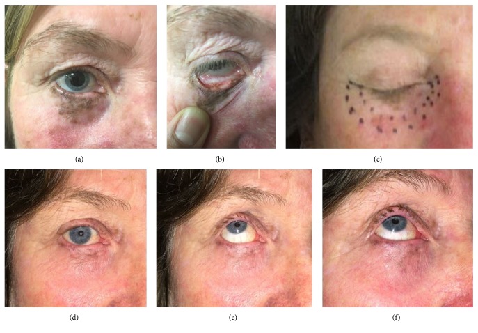
Figure 14.
(a, b) This lady had a significant pigmented lentigo maligna of the lower right eyelid. It spilt over onto the palpebral surface of the conjunctiva. (c) The next figure shows the CTV as marked by the radiation oncologist and then a 5 mm expansion to superficial radiotherapy field. (d, e, f) The following three images are photos taken six weeks after treatment. One can see significant resolution of the lentigo maligna and loss of the lower lid eyelashes but the globe is untouched. Eye and vision are fine as at baseline.

