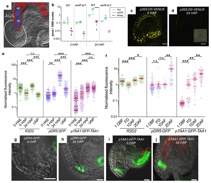
Fig. 1. Auxin accumulation in integuments.
a, Micropylar region of an Arabidopsis fertilized seed. Embryo and endosperm are colored in blue and red, respectively. ac, apical cell; bc, basal cell; C, chalaza; F, funiculus; o.i., outer integument; i.i., inner integument; the embryo attachment region is circled. b, Quantification of IAA, IAA-Aspartate (IAAsp), and oxidized IAA (oxIAA) in wild-type (WT) and wei8 tar1 ovules. c-d, p35S:DII-VENUS expression (yellow signal) in unfertilized (c) and 24 HAP (d) ovules. Inset in (d) shows the same ovule with enhanced brightness. e-f, Quantification in the embryo attachment region of GFP fluorescence (pDR5:GFP and pTAA1:GFP-TAA1) and mDII/DII ratio, normalized to 0 HAP (e) and FD (f). Data presented as individual points with a horizontal bar at the mean ± s.e.m at the vertical bar. g-j, pDR5:GFP and pTAA1:GFP-TAA1 expression in the wild-type embryo attachment region. Green GFP signal, merged with the brightfield image. Reddish signal, autofluorescence. Scale bars, 20 µm.

