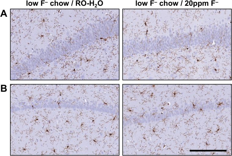
Fig. 7.
Representative images of Iba-1+ microglia in the hippocampus of adult Long-Evans hooded rats exposed to low-F− chow/RO-H2O or low-F− chow/20 ppm F− drinking water beginning on gestational day 6. (a) Suprapyramidal blade of the dentate gyrus. (b) CA1 pyramidal cell layer. Cells displayed normal process-bearing morphology with no evidence of reactivity or activation. 3,3-diaminobenzidine staining (brown). Hematoxylin counterstain (blue) (n = 6). Scale bar = 100 μm

