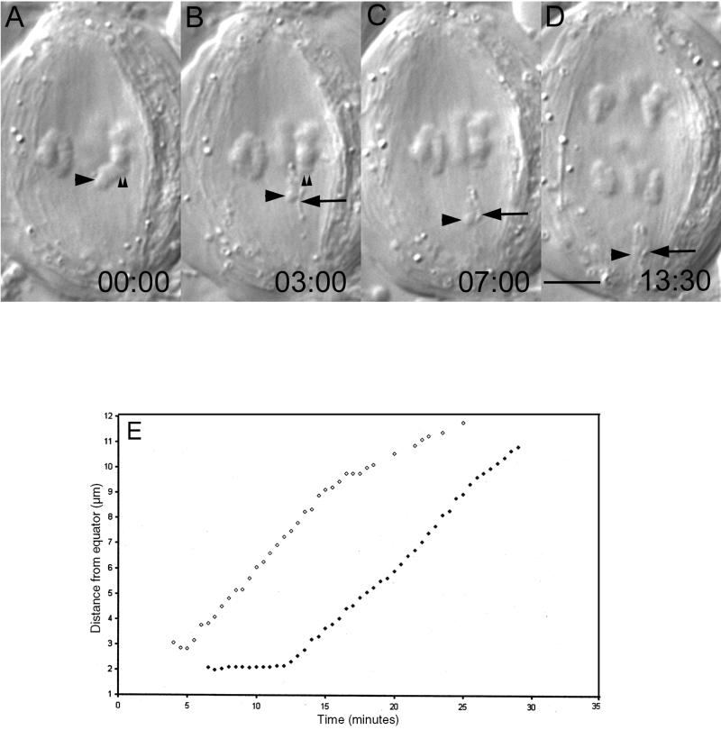
Figure 2.
Chromosome fragments lacking kinetochores are transported poleward. (A–D) Selected frames from a time-lapse recording of a primary (meiosis I) spermatocyte in which a pole-directed arm (A, large arrowhead) was severed (B) from the kinetochore region (A and B, small arrowheads) of a monochiasmic bivalent. This operation created a sniglet (B–D, large arrow) and an acentric chromosome fragment (B–D, large arrowhead). As in metaphase II (Figure 1), both the sniglet and acentric fragment moved poleward (D). Time in minutes:seconds. Bar (in D), 5 μm. (E) Distance from the equator versus time for the acentric fragment (○) depicted by the arrowhead in B–D, and also for the lower left half-bivalent (●) in D as it moved poleward during anaphase. Note that each moved with a similar kinetic profile.

