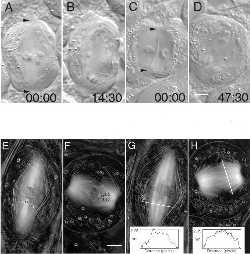
Figure 4.
Taxol treatment induces the formation of short, broad spindles, but taxol does not inhibit anaphase onset or prevent the subsequent poleward motion of chromosomes. (A–D) Differential interference contrast micrographs of living DMSO (0.05%) control (A and B) and taxol (5 μM for 15 min)-treated (C and D) spermatocytes during meiosis I at metaphase (A and C) and near the completion of anaphase A (B and D). Positions of the basal bodies of the polar flagella are marked with arrowheads in A and C. Note that anaphase takes ∼3 times longer in the shortened taxol spindle than in the DMSO control. Another noteworthy point, illustrated in C, is that mitochondria (normally restricted to the periphery of the spindle) often are found within the central domain of spindles formed in taxol. In this case, the mitochondrion appears as a highly refractile rod extending from pole to pole and between two bivalents. Time in minutes:seconds. Bar (in D), 5 μm. (E–H) Images generated with polarization microscopy depicting birefringence (bright contrast) due to spindle microtubules in untreated control (E and G) and taxol-treated (F and H) spermatocytes at metaphase. (E and F) Retardance magnitude was measured within areas 0.55 μm2; kinetochore fiber retardance appears reduced after 5 μM taxol treatment (2.0 vs. 2.35 nm in the control; refer to Table 2 for summary), but nonkinetochore domains appear less affected by taxol (1.5 vs. 1.62 nm in the control). (G and H) Similar results were obtained from line scans made perpendicular to the spindle axis. The top of the line scan in H (10 μM taxol) is on the left of the plot. Bar (in F), 5 μm.

