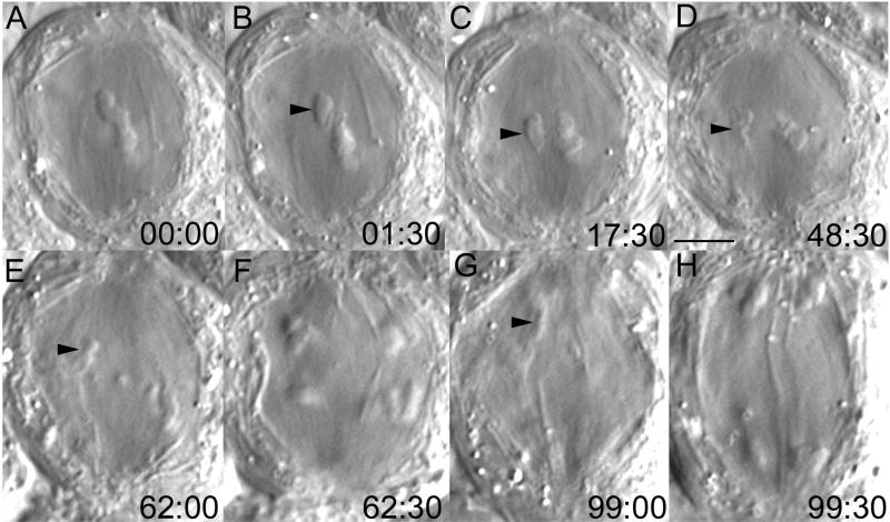
Figure 5.
Acentric fragments, generated near the spindle equator in taxol-treated spermatocytes, are not transported poleward as they are in control spermatocytes. (A–H) Selected frames from a time-lapse sequence of a taxol-treated primary spermatocyte that contained a pole-directed achiasmic arm (A–D, arrowhead). After it was severed with the laser (B), this arm remained relatively motionless over the next 47 min until it began to disintegrate before anaphase onset. (E–H) During anaphase, the fragment disintegrated further as it moved poleward (E and G), trailing behind the segregating half-bivalents (F and H), which moved poleward with a velocity of ∼ 0.1 μm/min. Time in minutes:seconds. Bar (in D), 5 μm.

