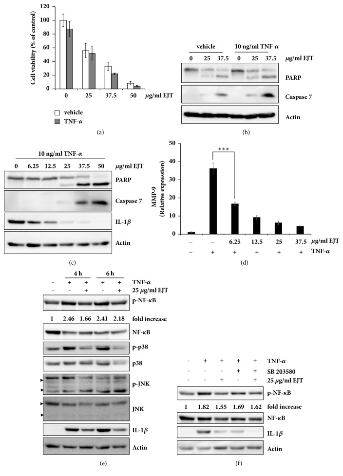
Figure 4.
EJT extract significantly decreased TNF-α-induced inflammation in MH7A cells. (a) MH7A cells were pretreated with 10 ng/ml TNF-α or DMSO for 30 min and then treated with the indicated concentrations of EJT extract for 24 h. The cell viability was determined by using an MTT assay. (b) Immunoblot analyses were performed by using specific antibodies. (c) MH7A cells were pretreated with 10 ng/ml TNF-α for 30 min and then treated with the indicated concentrations of EJT extract for 24 h. The cell lysates were subjected to SDS-PAGE and analyzed by immunoblotting with antibodies specific to PARP, caspase 7, IL-1β, and actin. (d) The relative mRNA level of MMP-9 was measured by using real-time quantitative PCR. Significant differences were indicated by ∗∗∗p < 0.001. (e) MH7A cells were pretreated with 10 ng/ml TNF-α for 30 min and then treated with 25 μg/ml EJT extract for indicated time. Immunoblot analyses were performed using specific antibodies. The fold increase in p-NF-κB expression is the ratio of the p-NF-κB to NF-κB. (f) MH7A cells were pretreated with 20 μM SB203580 for 2 h and then treated with 10 ng/ml TNF-α. After 30 min, MH7A cells were treated with 25 μg/ml EJT for 6 h. Immunoblot analyses were performed using specific antibodies. The fold increase in p-NF-κB expression is the ratio of the p-NF-κB to NF-κB.

