Fig. 6.
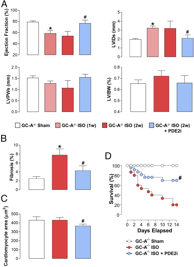
PDE2 inhibition reverses sympathetic hyperactivation-induced HF in GC-A−/− mice. Echocardiographic indices of heart structure and function (A), cardiac fibrosis (B), and cardiomyocyte size (C) are shown in sham mice and animals administered ISO (20 mg⋅kg−1⋅d−1 s.c.) for 2 wk (2w) in the absence and presence of BAY 60-7550 [10 mg⋅kg−1⋅d−1 p.o., initiated at 1 wk (1w)]. (D) Survival in sham mice and animals administered ISO (20 mg⋅kg−1⋅d−1 s.c.) for 2w in the absence and presence of BAY 60-7550 (10 mg⋅kg−1⋅d−1 p.o., initiated at day 0). Data are expressed as mean ± SEM and analyzed by one-way ANOVA with a Bonferroni post hoc test (A–C) or as a Kaplan–Meier survival plot (D). *P < 0.05 versus sham; #P < 0.05 versus AAC (6 wk; A–C) or GC-A−/− + ISO (D) (n = 6–12).

