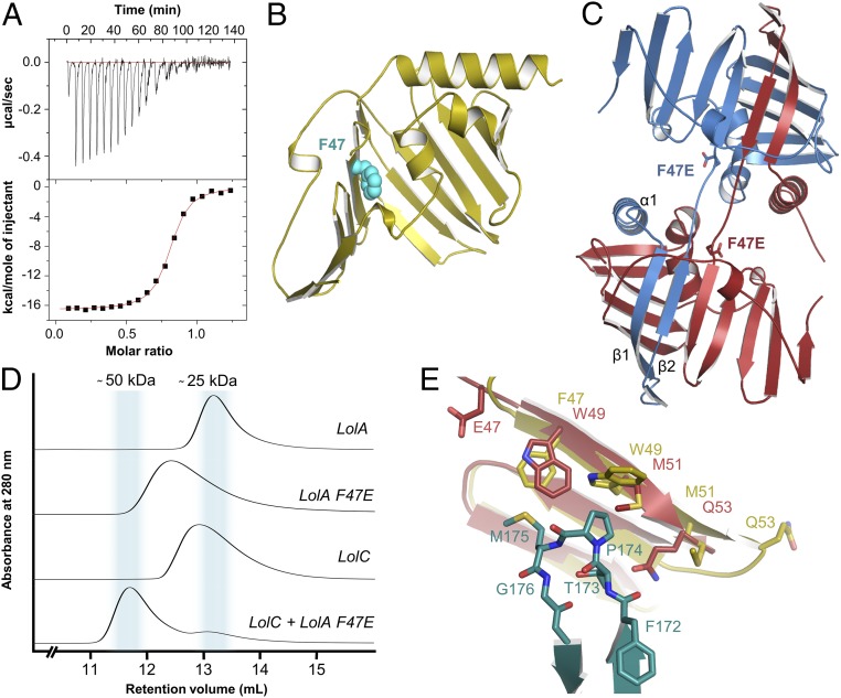
Fig. 5.
Structural and functional analysis of the tight-binding LolA F47E variant. (A) ITC experiment demonstrating binding of LolA F47E to the LolC periplasmic domain. (B) Location of residue F47 in wild-type LolA. (C) Crystal structure of LolA F47E revealing a domain-swapped dimer. (D) SEC experiment for wild-type and LolA F47E variant. (E) Close-up view of LolA F47E variant showing the strand-slip affecting the location of residues E/F47, W49, M51, and Q53. LolA wild-type and F47E are in yellow and red, respectively; LolC Hook is shown in teal.

