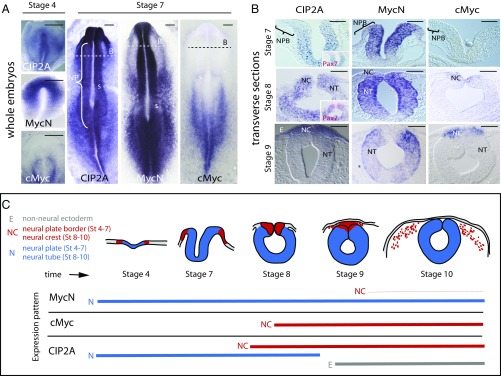
Fig. 1.
Expression patterns of CIP2A, MycN, and cMyc in the developing neural tube and neural crest cells in the chicken embryo. (A) Whole-mount in situ hybridization shows that CIP2A and MycN, but not cMyc, are expressed in the neural plate (NP) during gastrulation throughout the anterior-to-posterior axis. B, areas shown in panel B; s, somites. (B) In contrast, the neural plate border (NPB) that later forms the neural crest does not express any of the three genes at stages HH4–7 as also seen in transverse sections. As the neural tube closure begins at stage HH8, CIP2A is expressed throughout the entire neural tube (NT), whereas MycN is restricted to the ventral parts that will form the CNS. cMyc expression onsets at late stage HH8 in the dorsal neural tube that will become the neural crest (NC). By stage HH9 CIP2A and cMyc are restricted to the dorsal neural crest area, whereas MycN is expressed in the remaining neural tube but not in the dorsum. CIP2A expression is also seen in the nonneural ectoderm (E) after stage HH9. (Scale bars: 20 μm.) (C) A schematic diagram summarizing the expression patterns seen in A and B and SI Appendix, Fig. S1. Red represents the neural crest, blue represents neural stem cells of the future CNS, and gray is future epidermis. The thin red line for MycN represents a much lower expression level of MycN in the migrating neural crest compared with the other stages and genes, respectively.

