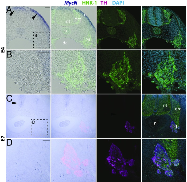
Fig. 2.
MycN is not expressed in the developing sympathetic ganglia. (A) At day 4 of chicken embryo development, MycN is expressed in the dorsal neural tube and the dermatome (arrowheads), but no MycN expression was detected in the peripheral ganglia as shown by in situ hybridization in the trunk level. The ganglia are highlighted by HNK immunostaining. (B) High-magnification images of the E4 sympathetic ganglia with no MycN expression. (C) At day 7, MycN expression is visible in the ventricular apical region surrounding the neural tube, but no expression is detected in the TH-immunopositive sympathetic ganglion. (D) High magnification of the E7 sympathetic ganglia with no MycN expression (boxed area in panel C). d, dermatome; da, dorsal aorta; drg, dorsal root ganglion; n, notochord; nt, neural tube; sg, sympathetic ganglion. (Scale bars: 100 μm.)

