Fig. 5.
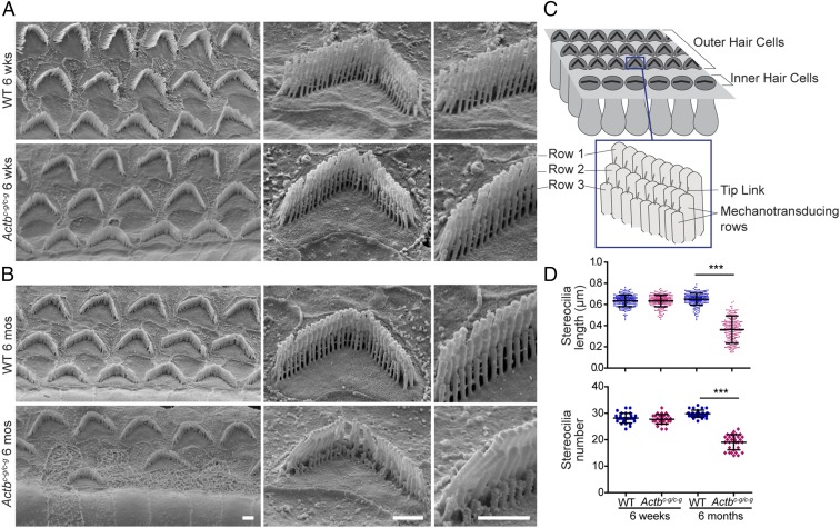
Actbc-g mice show evidence of stereocilia degeneration. Representative scanning electron microcopy images of (A) 6-wk-old and (B) 6-mo-old OHC stereocilia from the middle turn of the cochlea. (Scale bar: 1 µm.) (C) Cartoon image of the stereocilia. (D) Length (in micrometers) of stereocilia in row 3 of OHC bundles in 6-wk-old and 6-mo-old WT and homozygous Actbc-g mice. n = 4 mice, n ≥ 200 stereocilia. Number of stereocilia in row 3 of OHC bundles in 6-wk-old and 6-mo-old WT and homozygous Actbc-g mice. n = 4 mice, n ≥ 28 cells. One-way ANOVA with Tukey’s posttest was performed. ***P < 0.001. Error bars are SD.

