Fig. 4.
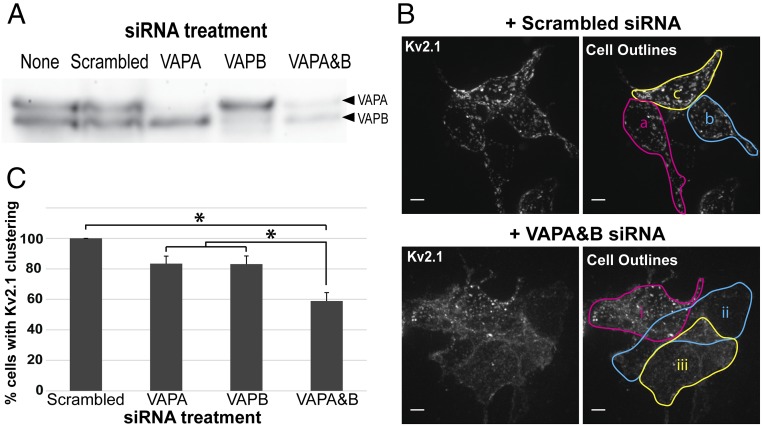
Effect of siRNA-mediated knockdown of VAPA and VAPB on Kv2.1 clustering. (A) Western blot demonstrating efficacy of VAPA and VAPB siRNA. Protein blot was probed with anti-VAPB antibody which cross-reacts with VAPA. (B) Representative image of GFP-Kv2.1-loopBAD clustering in the presence of scrambled or VAPA and VAPB siRNA taken via spinning-disk microscopy. z-stack maximum intensity projections are shown. In Upper Right image all three cells (a–c) were scored as having clustered Kv2.1. In the Lower Right image only cell i was scored as having clusters. Note that this image is presented simply to illustrate how clustering was defined as opposed to being quantitative with respect to the effect of the siRNA treatment. (Scale bars: 5 µm.) (C) Quantification of the percentage of cells displaying Kv2.1 clustering after various siRNA treatments. Eighty-six cells receiving the scrambled siRNA, 90 cells receiving VAPA siRNA, 61 cells receiving VAPB siRNA, and 144 cells receiving both VAPA and VAPB siRNA were examined within 26, 27, 21, and 41 images, respectively. Error bars represent SEM. For analysis, a one-way ANOVA was performed, F(3, 111) = 13.61, P = 1.27 × 10⁻⁷, with post hoc Tukey’s tests. *P < 0.01 significance.

