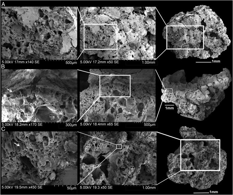
Fig. 3.
Scanning electron microscope images of bread-like remains from Shubayqa 1. (A) Sample number 6 showing the typical porous matrix of bread with small closed voids. (B) Detail of an aleurone layer from sample number 17 (at least single celled). (C) Sample number 12 showing vascular tissue, the arrow marks the xylem vessels in longitudinal section (for additional images of the remains see SI Appendix, Figs. S2–S8).

