Figure 5.
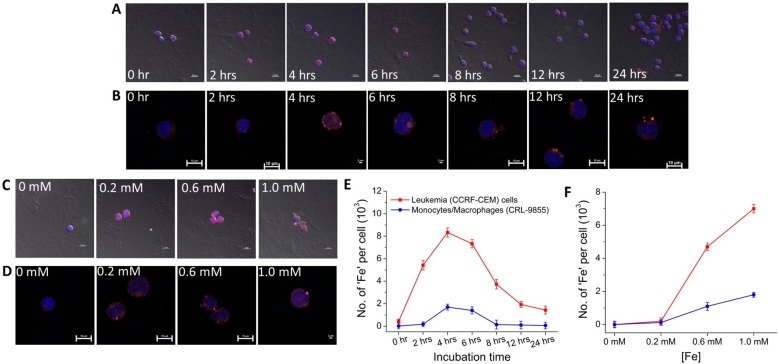
Fluorescence confocal microscopy (FCM) images and cellular uptake kinetics of the internalized ND-PEG-tNCIOs-(AF555Cdv) in (A) leukemia (CCRF-CEM) cells and (B) monocytes/macrophages (CRL-9855), incubated with 1 mM [Fe] for different incubation times (scale bar at 10 μm in panel A and at 2 μm for 4 and 6 h and the rest at 10 μm in panel B), and (C) leukemia (CCRF-CEM) cells and (D) monocytes/macrophages (CRL-9855), incubated with different [Fe] for 4 h (scale bar at 10 μm in panel C and scale bar at 2 μm for 1.0 mM and the rest at 10 μm in panel D). The Alexa Fluor 555 Cadaverine was adsorbed to ND-PEG-tNCIOs (bright orange); the nuclei were stained with DAPI (blue). (E) Cellular uptake as the function of incubation time. (F) Cellular uptake as the function of [Fe] in CCRF-CEM and CRL-9855.

