Figure 2.
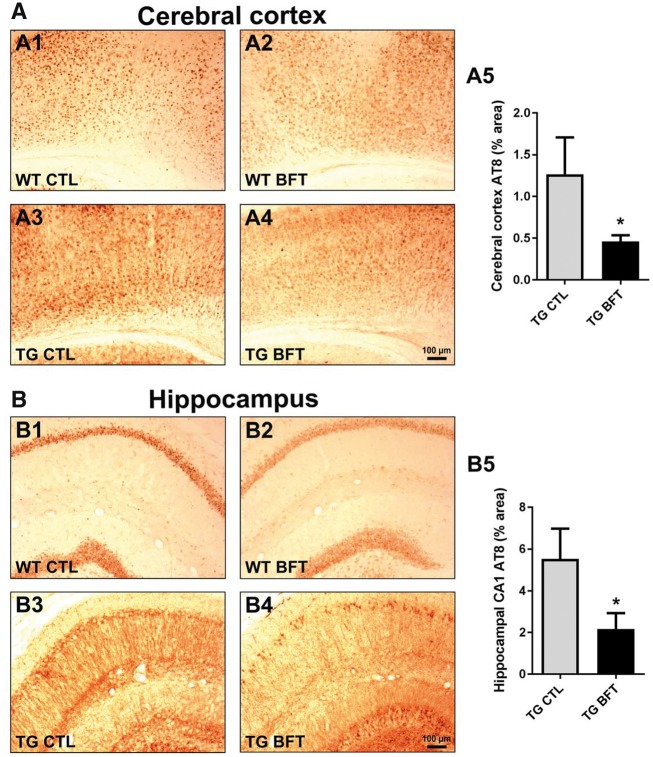
Reduced tau pathology following BFT administration. Immunostaining with AT8 antibody in the cerebral cortex (A) and hippocampus (B) of WT and TG mice treated with BFT or control diet. Calculation of the percent area occupied by AT8-immunopositive neurons revealed that BFT significantly reduced tau hyperphosphorylation in the brain of TG mice. For all tests, n = 6 for TG CTL and n = 8 for TG BFT. *P < 0.05 compared with TG CTL (unpaired t-test). Scale bar: 100 μm.

