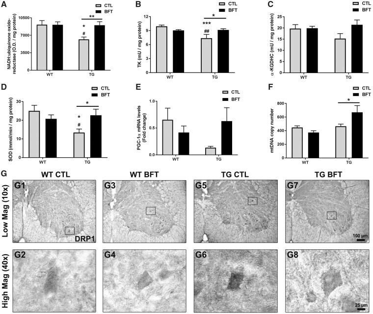
Figure 4.
BFT restores mitochondrial dysfunction. Frontal lobe samples were used to determine the mitochondrial expression of complex I (A), enzymatic activity of TK (B), α-KGDHC (C) and SOD (D), mRNA levels of PGC-1α (E), and mtDNA copy number (F). TG mice fed BFT exhibited a considerable increase in Complex I immunoreactivity, enzymatic activities, PGC-1α levels and the copy number of mtDNA. Data are mean ± S.E.M. of six to eight mice per group. ***P < 0.001 and *P < 0.05 relative to WT CTL. ##P < 0.01 and #P < 0.05 versus WT BFT. **P < 0.01 and *P < 0.05 compared with TG CTL (two-way ANOVA followed by Tukey multiple comparisons test). (G) Assessment of DRP1 immunoreactivity in the ventral horn of the lumbar spinal cord of WT and TG mice fed control or BFT diets. Insets correspond to high magnification images of DRP1 immunoreactivity in individual motor neurons. The expression of DRP1 was elevated in untreated P301S TG mice compared with WT mice with or without BFT. DRP1 immunoreactivity was robustly decreased by BFT treatment. Scale bar: 100 μm at low magnification and 25 μm at high magnification.

