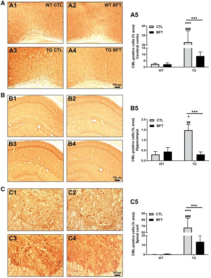
Figure 5.
Exposure to BFT prevents the formation of AGEs. CML immunoreactivity in the cerebral cortex (A), hippocampus (B) and spinal cord (C) of WT and TG mice with or without BFT treatment. There was a substantial decrease in the percent area occupied by CML-immunoreactive cells following BFT exposure in P301S TG mice. WT, n = 3–5; TG, n = 7–10. ***P < 0.001 and *P < 0.05 versus WT CTL. ###P < 0.001 and ##P < 0.01 compared with WT BFT. ***P < 0.001 relative to TG CTL (two-way ANOVA followed by Tukey multiple comparisons test). Scale bar: 100 μm.

