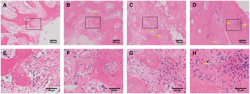
Figure 9.
Histopathological observations of bone regeneration at 12-week post-operation by HE staining. (A) and (E) for the BD group, (B) and (F) for the BD+ BMSCs group, (C) and (G) for the BD\CS + BMSCs group, and (D) and (H) for the BD\CS(pDNA-NELL1) + BMSCs group, yellow arrows for new vessels. (A–D) and (E–H) are different magnifications, and (E–H) are the amplification of the boxes in (A–D), respectively

