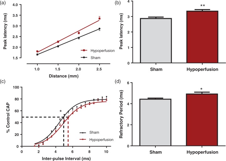
Figure 1.
Deficits in white matter function in response to severe chronic hypoperfusion in myelinated fibres. (a, b) There was a significant increase in peak latency at 2.5 mm from the stimulating electrode (**p = 0.003), indicative of slowed conduction of myelinated fibres, in hypoperfused animals. (c, d) For axonal refractoriness, the interpulse interval resulting in a 50% reduction in the CAP was significantly reduced in hypoperfused mice, indicative of perturbed axonal health. (*p = 0.03), when compared with sham-treated animals. Data are presented as mean ± S.E.M. Student’s t test, *p < 0.05 **p < 0.01, n = 12 per group.

