
Figure 2.
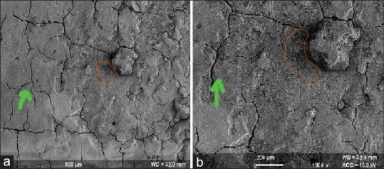
Test Group II (aided): Scanning electron microscope analysis of this group (×50 and ×100). It shows no visible smear layer, no residual embedded calculus, and slight loss of tooth substance with some roughness loss of the tooth surface. Loss of tooth substance is marked orange color with cracks seen with green marking. (a) Test Group II at 50x magnification; (b) Test Group II at 100x magnification; (orange colour) loss of tooth substance; (green colour) cracks on the root surface