
Figure 4. Redox state is perturbed by PFOS treatment, and modulated by Nrf2 deficiency.
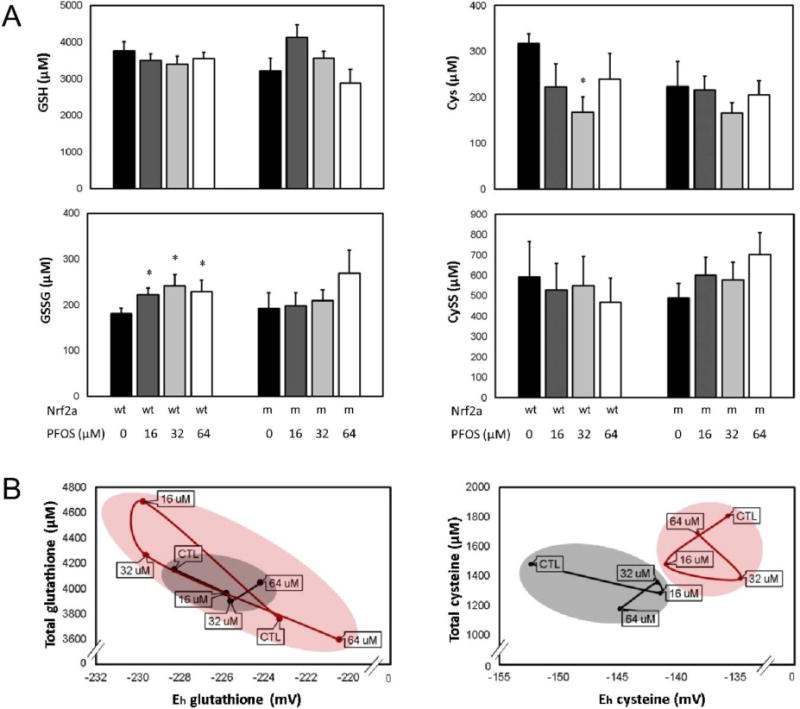
(A) Soluble thiol redox couples were quantified for the glutathione (GSH, GSSG; left) and cysteine (Cys, CySS; right) couples. PFOS increased oxidized GSSG and decreased Cys in wild-type (wt) embryos, though no effects were observed in mutants (m). (B) Total thiols and redox potential profiles were plotted to visualize the redox relationship between Nrf2a genotype and PFOS exposure for the glutathione (left) and cysteine (right) redox couples. In wild-type embryos (black), data were clustered more precisely and followed the expected trend—increasing PFOS exposures oxidized redox potentials. In Nrf2a mutants (red), two different profiles emerged for glutathione and cysteine. The total glutathione and glutathione redox potentials overlapped for wild-type and mutant embryos (left), though the range was much greater than in wild-type embryos. For cysteine (right), wild-type and mutant embryos had completely different profiles without overlap. Asterisks (*) indicate a dose-related change compared to genotypic controls. α = 0.05. n = 6-8 samples of 20 pooled embryos from 3-4 experimental replicates. Data presented are the mean and standard error of the mean (error bars in A, circle boundaries in B).