
Figure 5. PFOS increases apoptosis in wild-type embryos.
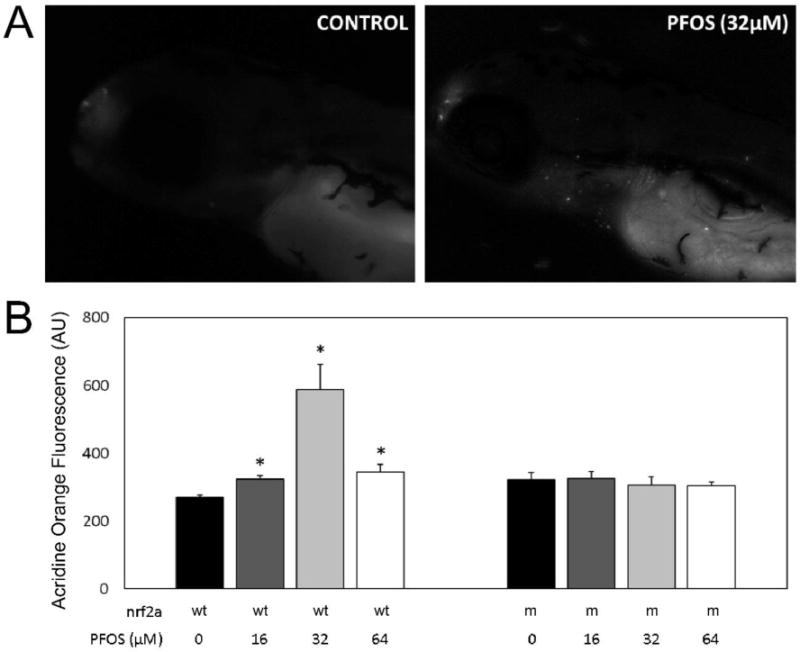
Acridine Orange staining was used to visualize apoptotic cells in vivo at 96 hpf in both wild-type (wt) and Nrf2a mutant (m) embryos. (A) Monochromatic epifluorescent images of control (left) and PFOS-treated (32 µM; right) embryos. An increased number of apoptotic cells can be seen in the rostrum, around the gills, and along the endodermal midline. (B) Homogenates of acridine orange stained embryos were used to quantitatively examine apoptosis. Nrf2a genotype for wild-type (+) and loss-of-function mutant (−) genotypes is provided below corresponding bars, as is PFOS exposure concentration. PFOS did not increase apoptosis in Nrf2a mutant embryos at any concentration. In wild-type embryos, PFOS increased apoptosis, with the greatest amount of apoptosis occurring in those exposed at 32 µM concentrations. Asterisks (*) indicate a dose-related change compared to genotypic controls. α = 0.05. n = 21-39 embryos from 3-4 experimental replicates