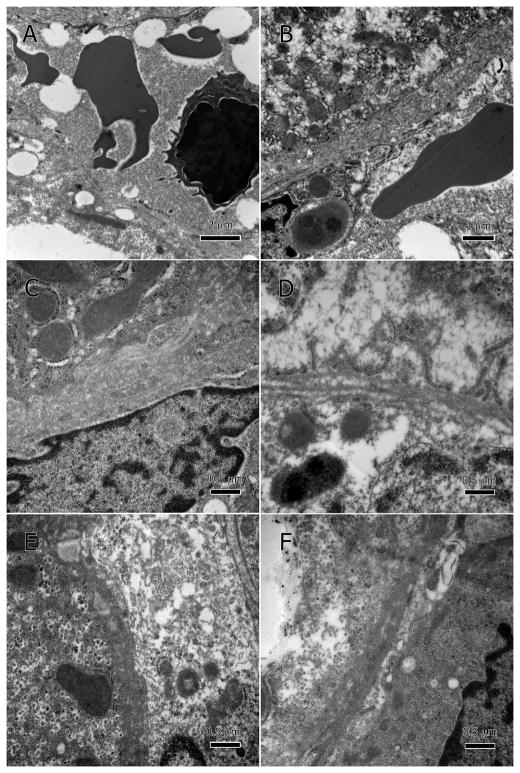
Figure 6.
Degenerate sinusoidal endothelium. Endothelial membranes show poorly defined margins, deterioration, and discontinuity; A and B at low magnification, and C–F at higher magnification. The degenerate endothelium has allowed hepatocytic cellular contents to leak into the sinusoidal lumen, especially large lipid droplets (A). Hepatocytes’ basal surface (adjacent to sinusoidal endothelium) lost its microvilli and the space of Disse was restricted or lacking.

