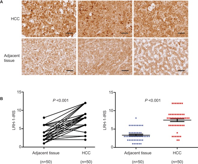
Figure 1.
LRH-1 was frequently upregulated in HCC clinical specimens.
Notes: (A) LRH-1 immunohistochemistry. Three representative micrographs of LRH-1-immunostained tissue microarray spots of HCC and adjacent tissues. Magnification, ×400; bars, 30 µm. The cells in tumor-adjacent tissues showed detectable or weak nuclear immunoreactivity. Intense nuclear immunosignals were detected in cancer cells in HCC tissues. (B) LRH-1 IRS analysis performed on HCC and adjacent tissues. Results showed that HCC tissues showed significantly higher LRH-1 expression than tumor-adjacent tissues. P<0.001 vs tumor-adjacent tissues.
Abbreviations: HCC, hepatocellular carcinoma; IRS, immunoreactivity score.

