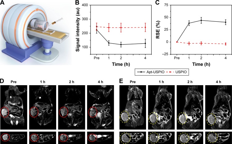
Figure 8.
In vivo MRI of xenograft tumors. (A) In vivo T2-weighted MRI of the HCC xenograft models after injection of USPIO or Apt-USPIO. (B) T2-weighted MR signal intensities of the Huh-7 tumors significantly decrease in the experimental (Apt-USPIO) group as compared to the control (USPIO) group. (C) RSE values in the experimental group are significantly higher than the control group at different time points. (D) Coronal and axial T2-weighted MR images of tumor-bearing mice before and after intravenous injection show significant negative enhancement of the tumors (red circles) at different time points in the Apt-USPIO group. (E) There is no decline in the signal intensities of the tumors (yellow circles) in the corresponding USPIO group.
Abbreviations: Apt-USPIO, aptamer-mediated USPIO; HCC, hepatocellular carcinoma; MRI, magnetic resonance imaging; RSE, relative signal enhancement; USPIO, ultrasmall superparamagnetic iron oxide.

