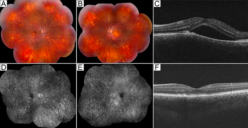
Figure 3:

Posterior uveitis in a 21-year-old male with ALPS-U (ALPS with undetermined genetic defect). (A,B) Color fundus photographs show scattered deep white choroidal lesions in both eyes (A, right eye; B, left eye). (C) Optical coherence tomography shows submacular fluid in the right eye. (D,E) Fluorescein angiography shows staining of deep choroidal lesions in both eyes and mild perifoveal leakage in the left eye (D, right eye; E, left eye). (F) With treatment, the patient’s chorioretinal lesions (not shown) and submacular fluid resolved.
