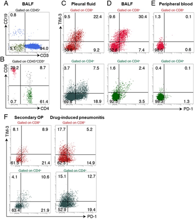
Figure 2. Flow cytometric analysis of lymphocyte subsets in BALF, pleural fluid, and peripheral blood of the patient.
(A, B) Expression of CD19 and CD3 on CD45+ cells (A) and expression of CD4 and CD8 on CD45+ CD3+ cells (B) in BALF. (C–E) Expression of PD-1 and TIM-3 on CD8+ T cells and CD4+ T cells in pleural fluid (C), BALF (D), and peripheral blood (E). (F) Expression of PD-1 and TIM-3 on CD8+ T cells and CD4+ T cells in BALF of patients with secondary organizing pneumonia (OP) or drug-induced pneumonitis.

