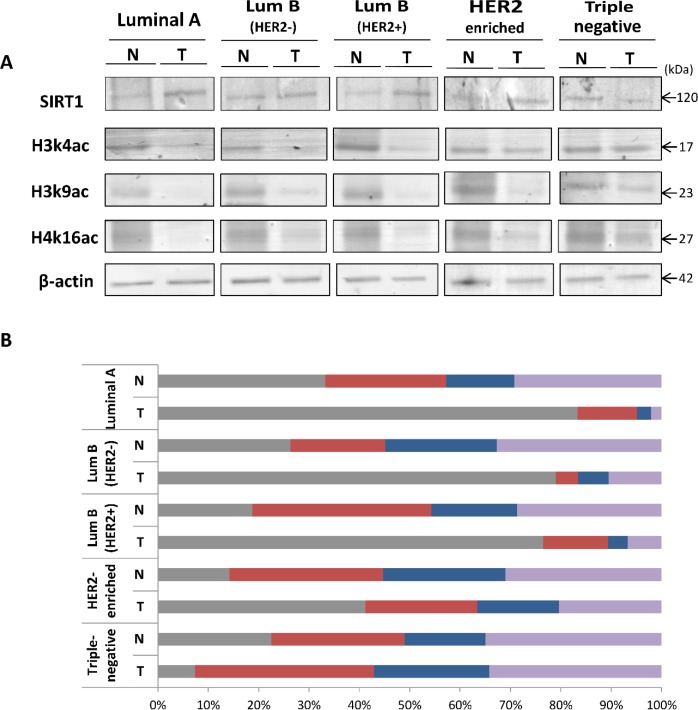
Figure 1. Differential expression patterns of SIRT1, H3k4ac, H3k9ac and H4k16ac in the 5 molecular breast tumor subtypes compared to matched normal tissues.
(A) Equal amounts of proteins were immunoblotted with anti-SIRT1 Ab (120 kDa), anti-H3k4ac Ab (17 kDa), anti-H3k9ac Ab (23 kDa) and anti-H4k16ac Ab (27 kDa). β-actin (42 kDa) served as an internal loading control. (B) Relative expression levels were evaluated using Quantity One software and normalized against the internal control β-actin. Each bar represents the percentage contribution of each of the 4 proteins compared to the total set as (100%). All experiments were performed in triplicate fashion. N: Normal, T: Tumor.

