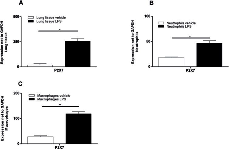
Figure 3. Significant upregulation of P2X7R-expression in lung tissue and BALF cells after LPS exposure.
Animals received LPS and vehicle on day 0 and were killed 24 hours later. Lung tissue and BAL-cells were collected and RNA was isolated. Relative expression of the P2X7R compared with GAPDH was analyzed using quantitative RT-PCR. (A) P2X7R subtype expression in lung tissue of PBS-exposed or LPS-exposed animals. (B) Expression of P2X7R on pooled BALF neutrophils (n = 10 LPS animals and n = 20 PBS animals). (C) P2X7R Expression on pooled BAL macrophages (n = 10 LPS animals and n = 20 PBS animals). Data are means ± SEM, n = 10–20, *p < 0.05; **p < 0.01; ***p < 0.

