
Figure 1.
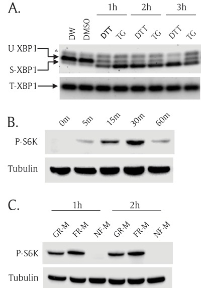
Both ER stress and TOR pathways are functional in S2R+ Drosophila cells. (A) ER stress inducers activated XBP1 mRNA splicing in S2R+ Drosophila cells. S2R+ cells were incubated with ER stress inducers for 1 hour, 2 hours, and 3 hours; then, total RNA was extracted and used to generate cDNA. Unspliced XBP1(U-XBP1) and spliced XBP1 (S-XBP1) were amplified using specific primer set across the XBP1 splicing region. Total XBP1 (T-XBP1) was used as a loading control. Asterisk (*) indicates a non-specific band. 1 mM dithiothreitol (DTT) and 0.2 μM thapsigargin (TG). Distilled water (DW) is the vehicle for DTT and Dimethylsulfoxide (DMSO) is the vehicle for TG. (B) Insulin increased phosphorylation of S6K. S2R+ cells were incubated with insulin (10 mg/ml) for multiple incubation times: 0 minutes, 5 minutes, 15 minutes, 30 minutes, and 60 minutes. The cell lysates were subjected to SDS-PAGE and transferred to nitrocellulose paper followed by Western blot analysis. Phospho(Thr398)-specific S6K antibody (P-S6K) detects only phosphorylated form of S6K. Tubulin serves as a loading control. (C) Nutrient-free (NF-M) media completely abolished S6K phosphorylation. S2R+ cells were cultured in growth media (GR-M) for 1 day. Then the cells were incubated with fresh media (FR-M) or nutrient free-media (NF-M) for 1 hour and 2 hours. Phospho(Thr398)-S6K and tubulin were detected by Western blot analysis.