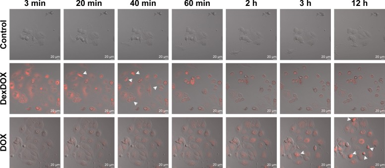
Figure 3.
Cellular penetration and subcellular distribution of DexDOX.
Notes: Representative images from video-assisted confocal microscopy showed that DexDOX entered MCF-7 more rapidly than DOX. The fluorescence signal was rapidly lost without entering the nucleus. Interestingly, pyknotic cells, which are indicative of apoptosis, were observed (arrow) after the fluorescence signal was lost. In contrast, DOX gradually diffused into cells and accumulated in the nucleus. The apoptotic cells (arrow) were observed in the later phase.
Abbreviation: DOX, doxorubicin.

