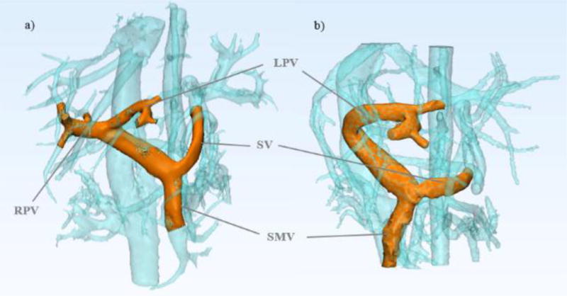
Figure 1.

4D flow MRI can be used to depict in vivo anatomy before and after partial hepatectomy in healthy living donors. The models shown were created from MR image data for a) Pre-surgery donor 1 portal vasculature and b) Post-surgery donor 1 portal vasculature.
