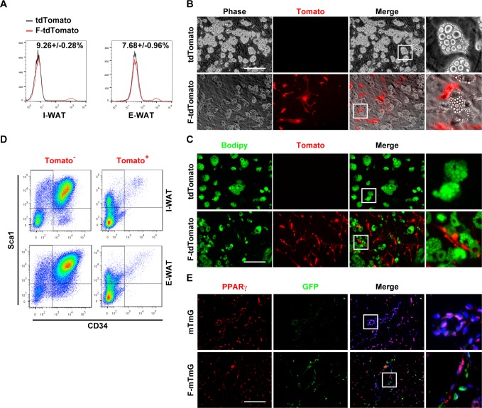
Fig 1. FSP1+ fibroblasts localize adjacent to preadipocytes without adipogenic potential.
(A) FACS analyses of tdTomato+ cells in SVF cells isolated from I-WAT and E-WAT of 4-month-old Rosa26-tdTomato (mT) and Fsp1-Cre; tdTomato (F-mT) mice. (B, C) SVF cells isolated from Rosa26-tdTomato (mT) and Fsp1-Cre; tdTomato (F-mT) I-WAT were adipogenically induced. Cells were stained with Bodipy 493/503 (panel C). Scale bar: 200 μm. (D) FACS analyses of CD34+Sca1+ cells in I-WAT and E-WAT SVF of 4-month-old Fsp1-Cre; tdTomato mice. (E) Immunofluorescent staining of GFP and PPARγ on I-WAT sections of 4-month-old mTmG and Fsp1-Cre; mTmG (F-mTmG) mice. Scale bar: 200 μm. CD34, cluster of differentiation 34; E-WAT, epididymal white adipose tissue; FACS, fluorescence-activated cell sorting; FSP1, fibroblast-specific protein-1; GFP, green fluorescent protein; I-WAT, inguinal white adipose tissue; PPARγ, peroxisome proliferator-activated receptor-γ; Sca1, stem cell antigen 1; SVF, stromal vascular fraction.

